Abnormal Shape
ear surgery centerProminent ear
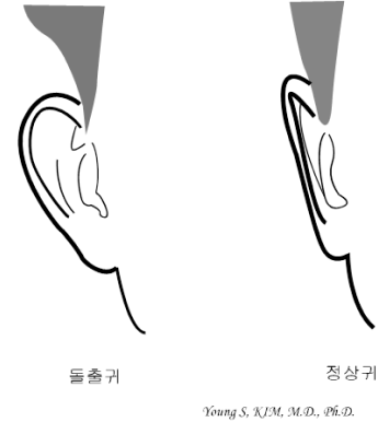
Prominent ear
Prominent ears are the most common congenital deformity of the ears,
and are also called donkey ears or Mickey Mouse ears.
The ears are far away from the head, and when viewed from the front,
the ears look spread out.
Since it occurs for various reasons, accurate correction
according to the cause is required to make natural ears.
In general, the unformed antihelix of the ear or hypertrophy of the auricular
concha are known to be the main causes of prominent ears,
but there are many other causes.
Experientially, prominent ears can be classified into
about 5 types depending on the cause,
and correction according to
these
classifications can produce more accurate and natural results.
Classification of Prominent ear (Bona Clinic's own taxonomy)
- Type Ⅰ
- Usually, it is a case where a large wheel is not formed, and it can be classified into two types in detail.
- Type Ia
- In the most common form, when the entire large two wheel is not formed except for the lower angle of the large two wheel.
-
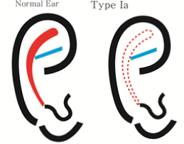
example picture
-
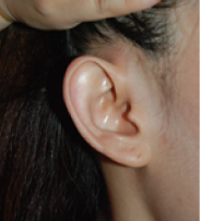
example photo
-
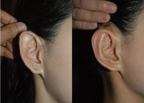
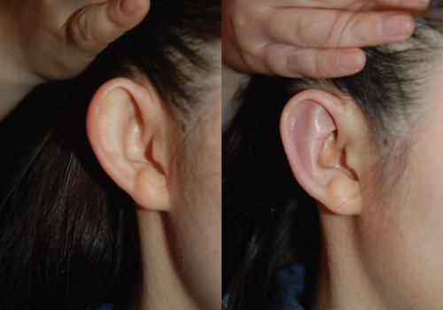
before and after surgery
- Type Ib
- If the formation of the lower angle including the amortization of the large two wheels is insufficient
-
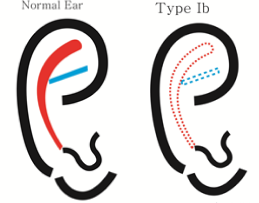
example picture
-
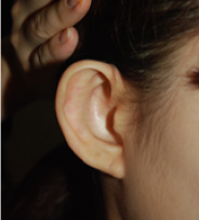
example photo
-
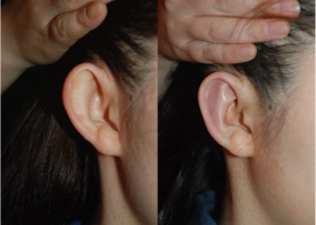
before and after surgery
- Type II
- In case of hypertrophy of the concha
-
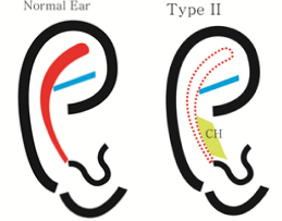
example picture
-
before and after surgery
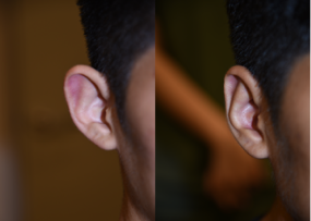
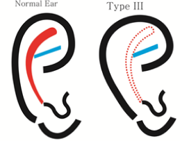
- Type III
- In case of helix-scapha hypertrophy
-
example picture
-
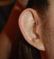
example photo
-
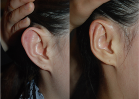
before and after surgery
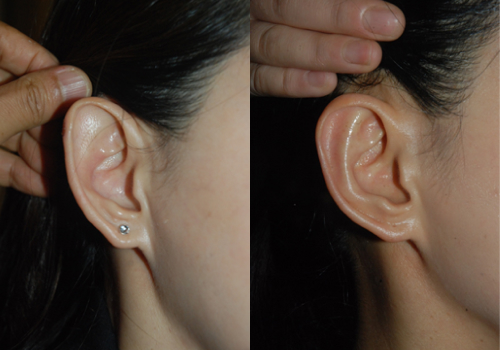
- Type IV
- If more than one variant exists
-
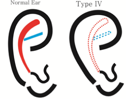
example picture
-
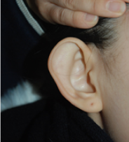
example photo
-
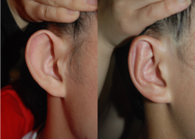
before and after surgery
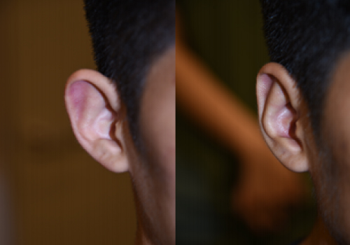
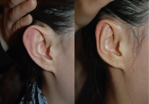
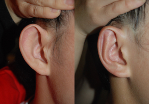
Before & After
-
 BeforeAfter
 -
 BeforeAfter
 -
 BeforeAfter
 -
 BeforeAfter
 -
 BeforeAfter
Consult with Bona Clinic.
information
-
Weekdays
10:00 ~ 19:00 Lunch time 13:00 ~ 14:00 -
Saturday
10:00 ~ 16:00 Lunch time 13:00 ~ 14:00 -
Directions
Line 3 Apgujeong
Station Exit 4 150m straight
(Line 3 Apgujeong Station Exit 4, 150m straight) Business registration. 211-09-47985
Copyright © 2023 Bona Clinic All RIGHTS RESERVED.